
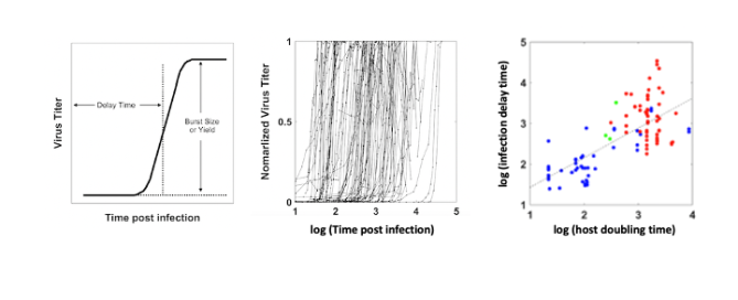
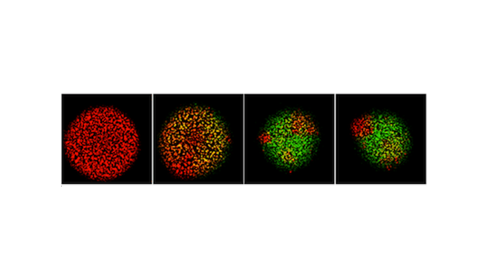
How does the timing of virus infection depend on their host?
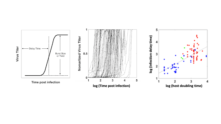
To address this question, consider the delay time that describes how long it will take, following the start of infection, for virus progeny to be released from infected cells (left). Analysis of such growth curves for more than 100 viruses indicate delay times from less than 100 to more than 10,000 minutes or about 1
